> Large size
|
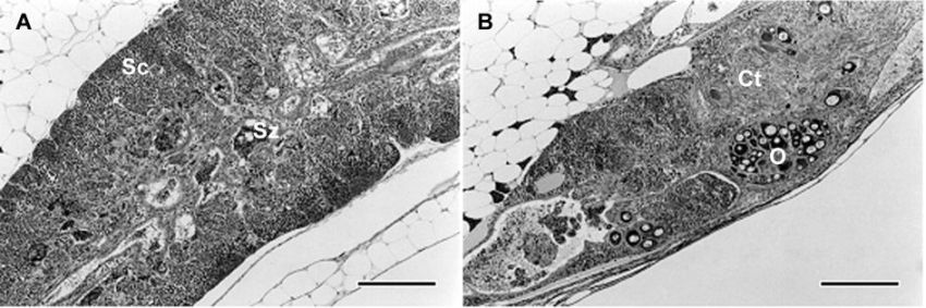
Figure 8-4. Gonadal sections (3 μm) from Japanese medaka at 60-dph in the parent generation of the fish full life cycle test, stained with hematoxylin and eosin. Each bar represents 400 μm. (A) Testis of a control male, showing normal spermatogenesis: spermatocytes (Sc) and spermatozoa (Sz). (B) A progressed testis-ova of a male exposed to 27.9 ng/L of 17β-estradiol. Dozens of oocytes (O) appear in the testicular tissue, accompanying abnormal connective tissue (Ct) with fewer testicular germ cells. (Modified from Seki et al., 2005. Copyright (2005) Environmental Toxicology & Chemistry, Allen Press Publishing Services.) |