|
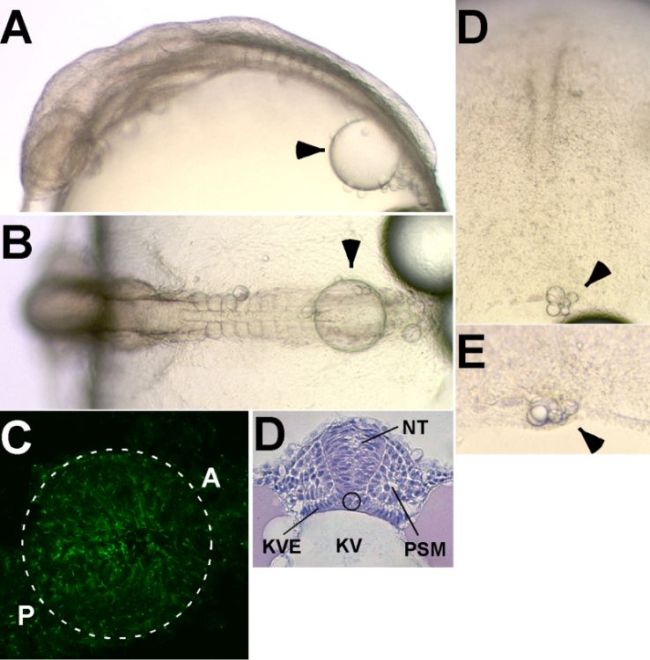
Figure 6-70.
Kupffer’s vesicle. (A) Lateral view at stage 22. (B) Dorsal view at stage 22. (C) Dorsal view at stage 16/17. (D) Lateral view at stage 16/17. (E) Monocilia in Kupffer’s vesicle, stained with antiacetylated-α-tubulin antibody. (F) Cross section of Kupffer’s vesicle. NT, neural tube; NC, notochord; KV, Kupffer’s vesicle; KVE, KV epithelium; PSM, pre-somitic mesoderm. KV is indicated by an arrowhead in A–D. (Reproduced from Hoji et al., 2007, with permission of John Wiley & Sons, Inc.; S. Takashima, unpublished data) |