|
|
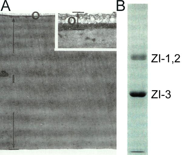
Figure 6-62.
(A) Transmission electron micrograph of sectioned 1-dpf egg envelope. The inset shows a magnified region of part of the outer layer. I, inner layer; O, outer layer. (Yamamoto and Yamagami, 1975) (B) SDS-PAGE pattern of component glycoproteins of the inner layer of the unfertilized egg envelope. (Hamazaki et al., 1987. Reproduced with permission of John Wiley & Sons, Inc.) |