|
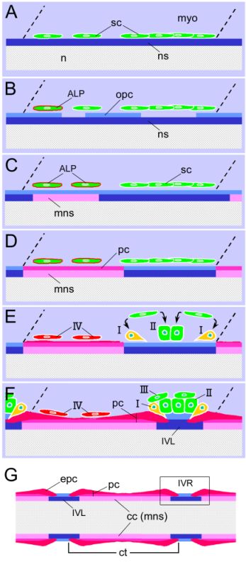
Figure 6-55.
Schematic diagram showing the formation of the medaka vertebral column. (A–F) Horizontal views of the right side of a segmental component. Anterior is to the left. Dotted lines show the segmental boundary of the myotome. (A) The embryo before 4-dpf. (B) 4-dpf embryo. (C) 5-dpf embryo. (D) Newly hatched larva (6-dpf). (E) After 3-dph. (F) Juvenile larva. (G) Schematic diagram of the larval vertebral structure. The diagram shows a horizontal view of a centrum and portions of the adjoining anterior and posterior centra. The box indicates the intervertebral region (IVR), which includes an intervertebral ligament and the bone edges of the two adjoining centra. sc, sclerotomal cell; ALP, ALP-positive sclerotomal cell; ct, centrum; epc, bone edge of perichordal centrum; IVL, intervertebral ligament; mns, mineralized notochordal sheath; myo, myotome; n, notochord; ns, notochordal sheath; opc, osteoid of perichordal centrum; pc, ossified perichordal centrum; I, type I cells, which are responsible for bone matrix formation of the perichordal centrum and mineralization on the edges of the centrum; II, type II cells, which are involved in the deposition of the collagenous matrix of the extra elastica; III, type III cells, which are adjacent to both the type I cells and the type II cells, but not directly facing the bone matrix of the centrum; IV, type IV cells, which are responsible for bone matrix formation and mineralization of the middle part of the perichordal centrum. (Inohaya et al., 2007. Reproduced with permission of John Wiley & Sons, Inc.) |