|
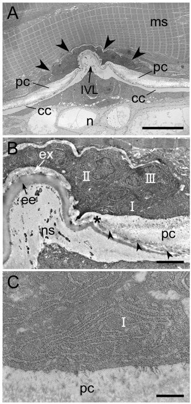
Figure 6-54.
Transmission electron micrographs showing horizontal histological sections of the intervertebral region of growing medaka larvae (body length of approximately 7 mm). Anterior is to the left. (A) Low-magnification micrograph showing the intervertebral region. Electron-dense cells are observed in the intervertebral region (arrowheads). (B) Magnified region of the image shown in A. The intervertebral ligament is composed of three matrix layers: the extra elastica (ex), the elastic externa (ee), and the notochordal sheath (ns). The fibrous layer of the extra elastica also constitutes the innermost region of the perichordal centrum (arrowheads). Cuboidal- or oval-shaped cells are directly located on the outer surface of the extra elastica and on the bone edge of the perichordal centrum (pc). These cells are classified into three types (I–III; see text for further details). (C) High-magnification micrograph of a type I cell seen in B. Many rough endoplasmic reticula (RER) can be seen in the cytoplasm of the type I cell. cc, chordal centrum (mineralized notochordal sheath); IVL, intervertebral ligament; ms, muscle; n, notochord; pc, perichordal centrum. The asterisk in B shows the edge of the perichordal centrum adjacent to the intervertebral ligament. Scale bars: A, 10 μm; B, 2 μm; C, 500 nm. (Inohaya et al., 2007. Reproduced with permission of John Wiley & Sons, Inc.) |