|
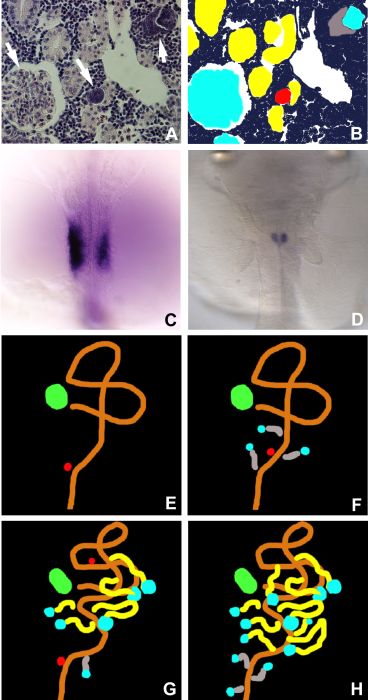
Figure 6-45A,B > Large size
Figure 6-45.
(A) Histological section of the mesonephros, showing developing nephrons at three different stages of development. Middle arrow, mesenchymal condensate; right arrow, nephrogenic body; left arrow, glomerulus of the mature nephron. (B) Schematic illustration of A; the colors correspond to those in (E, F, G, and H). Red, mesenchymal condensate; light blue, glomerulus; grey, immature tubule without a lumen, the tail portion of the nephrogenic body; yellow, mature tubule connecting to the pronephric tubule or duct; purple, mesenchymal cells. (C, D) WT1 expression at stage 21 (C) and at stage 33 (D). Illustration of nephron formation at 5-dph (E), 7-dph (F), 10-dph (G), and 15-dph (H). Green, pronephric glomerulus; orange, pronephric tubule and duct; other colors are the same as for B. (C, D) Dorsal views. |