|
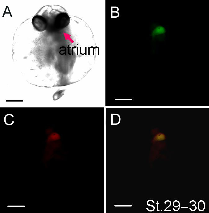
Figure 6-42.
Whole-mount immunohistochemical detection of the chamber structure of the developing heart using MF20 (monoclonal anti-chick myosin antibody) andS46 (monoclonal anti-myosin antibody). (A) Light field image of an embryo at stage 29–30. Immunoreactivity of the developing heart chamber against (B) S46 and (C) MF20. (D) Superimposed image of b and c. Scale bar, 250 μm. |