|
|
|
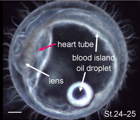
Figure 6-36.
Left side lateral view of a stage 24–25 embryo. Heart tube development continues (see also Movie M6-2b). Scale bar, 250 μm. |
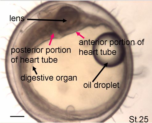
Figure 6-37.
Right side lateral view of a stage 25 embryo. The heart beats are observed as two motions near the anterior and posterior portions of the eye vesicle. Scale bar, 250 μm. A countercurrent of the blood stream can be seen in Movie M6-2c. |