|
|
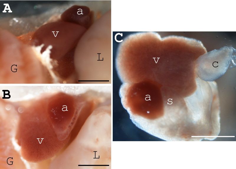
Figure 6-30. The mature medaka heart as observed using a dissecting microscope. After fixation with 4% paraformaldehyde, the body was opened carefully (A and B show different angles), and the whole heart was dissected from the body (C). a, atrium; G, gill; L, liver; V, ventricle; c, conus arteriosus; s, sinus venosus. Scale bars, 0.5 mm. |