|
|
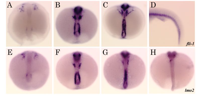
Figure 6-26. Expression patterns of hemangioblasts and vascular endothelial cell markers. (A–D) fli-1 expression at stages 18 (A), 20 (B), 23 (C), and 28 (D). (E–H) lmo2 expression at stages 18 (E), 20 (F), 23 (G), and 25 (H). (A–C, E–H) Dorsal view. Rostral is to the top. (D) Lateral view. Rostral is to the left. |