|
|
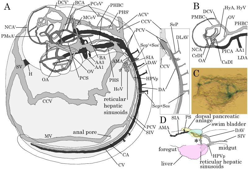
Figure 6-24. Circulation in the developing medaka at stage 29. (A) Diagram of the embryonic circulatory system. Dorsolateral view. (B) Diagram of the vessels around the left eye. Ventrolateral view. (C) The dissected digestive anlages of a Berlin blue dye-injected specimen showing the hepatic portal system at the boundary between the foregut and midgut. Dorsolateral view. (D) Diagram of C. Rostral is to the left in all panels. The names of major anlages are noted in D as landmarks. (Fujita et al., 2006. Reproduced with permission of John Wiley & Sons, Inc.) |