|
|
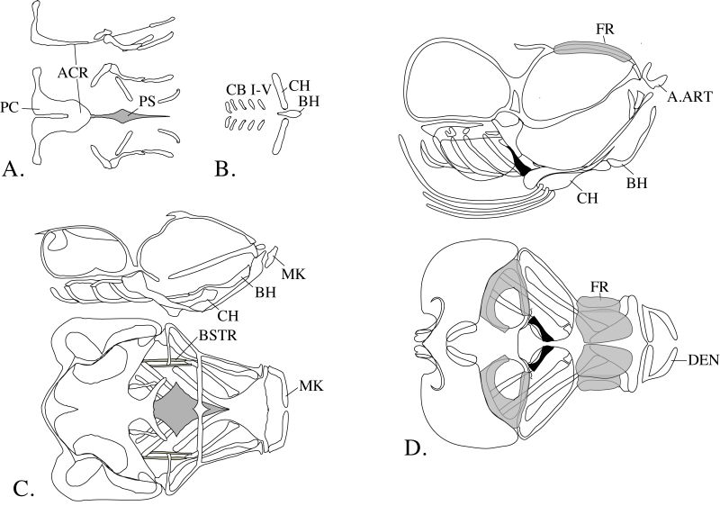
Figure 6-18. Diagrams of cranial and visceral arch skeletons from stage 33 (A, B), 40 (C), and 43 (D) embryos. In all figures, rostral is to the right. In A, C, and D, the lateral view is shown at the top and the dorsal view at the bottom. ACR, acrochordals; A.ART, anguloarticular bones; BH, basihyal; BSTR, branchiostegal; CB, ceratobranchial; CH, ceratohyal; DEN, dentaries; FR, frontal; MK, Meckel’s cartilage; PC, parachordal; PS, parasphenoid. (Modified from Langille and Hall, 1987, with permission of John Wiley & Sons, Inc.) |