|
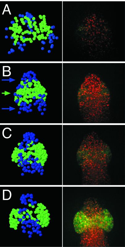
Figure 6-16. Morphogenesis of the optic vesicle. A The eye field, highlighted by the expression of rx3::GFP, is established within the anterior neural plate. B Upon formation of the neural keel, rx3-positive retinal progenitor cells are modulated in their approach toward the midline, priming the evagination site. C Epithelialization forms a neck posterior to the prospective optic vesicles and spreads in the posterior direction. D The optic vesicles evaginate due to the migration of individual cells that delaminate from central positions and intercalate into the forming optic bud. Images on the left are reconstructions from cell tracking data, and those on the right are right half central Z-sections through the forebrain/eye region. All images show the dorsal view, with anterior at the top. Nuclei are labeled using histone-dsRed fusion proteins and retinal progenitor cells are marked using rx3::GFP. |