|
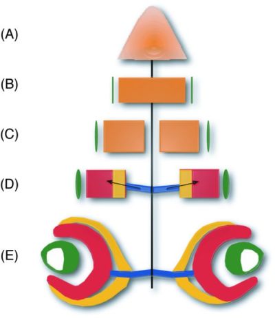
Figure 6-15. Schematic representation of eye development. The central line shows the temporal axis and also the midline of the developing embryo. (A) Eye development initiates during gastrulation by specification of the anterior neural plate. The tissue from which the neuroectodermal part of the eye will form (retina and optic stalk) is shown in orange. (B) By the end of gastrulation, the eye field is determined and the retinal identity is established. Tissue that will give rise to the lens is shown in green. (C) In response to signals emanating from the midline, the initially single retinal anlage is split into two retinal primordia. (D) Evagination of the optic vesicle occurs via active morphogenesis and proliferation, and then the optic vesicles invaginate to form the optic cup. The neural retina (dark orange), pigmented epithelium (pale orange), and optic stalk (blue) form. The arrow indicates the secreted signals that initiate retinal differentiation. (E) The neural retina differentiates and sends axons along the optic stalk to the primary targets in the optic tectum. At this stage of eye development, the differentiated lens is fully embedded into the optic cup. (Adapted from Wittbrodt et al., 2002.) |