|
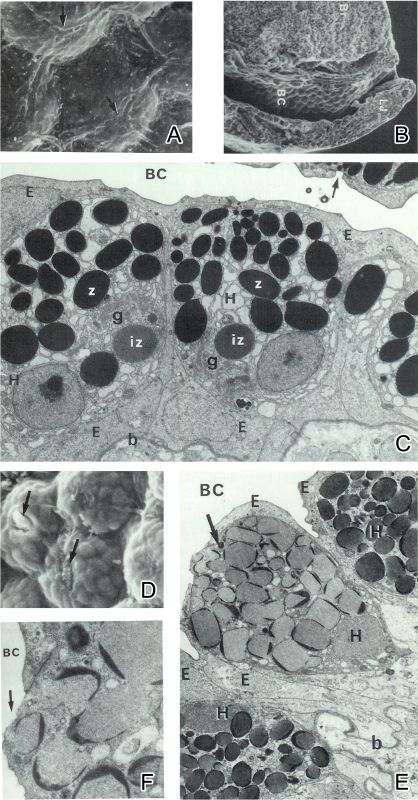
Figure 6-14. Ultrastructural changes in the hatching gland cell during secretion of hatching enzyme. A Scanning electron micrograph of the surface epithelial cells covering the hatching gland cells. Three adjoining cells meet (arrow) at the apical center of an underlying gland cell. B Scanning electron micrograph of a median section of the head of a prehatching embryo. Br, brain; BC, buccal cavity; LJ, lower jaw. C Transmission electron micrograph of the hatching gland cells of a prehatching embryo. g, Golgi complex; z, zymogen granules; iz, immature zymogen granules; b, basal lamina; E, epithelial cells. D Scanning electron micrograph showing the swelling of the hatching gland cells. Some epithelial joints are separated (arrow). E Transmission electron micrograph of the hatching gland cells in the process of secretion (arrow). H, hatching gland cells. F Transmission electron micrograph of the apical portion of hatching gland cells. Cytoplasm mixed with secretory substances is flowing out into the buccal cavity (arrow). From Yamamoto et al. (1979). |