|
|
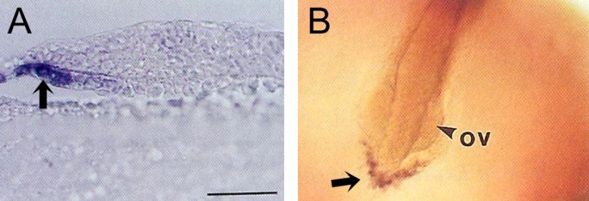
Figure 6-12. Early expression of hatching enzyme genes. (A) Median section of a stage 16–17 embryo hybridized with a high choriolytic enzyme probe. Several cells located at the anterior part of hypoblast layer were stained (arrow). Scale bar, 50 μm. (B) Whole mount in situ hybridization of a stage 19–20 embryo (3–4 somites). Anti-sense RNA of high choriolytic enzyme cDNA was used as a probe. V-shaped cell mass located in front of the embryonic body was stained (arrow). ov, optic vesicle. From Inohaya et al. (1995). |