|
|
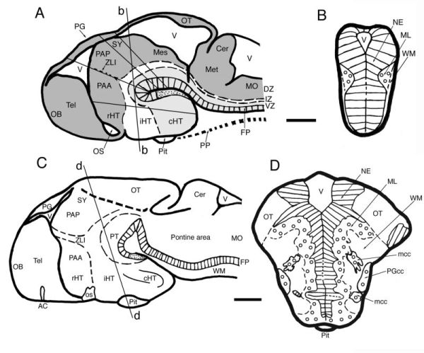
Figure 6-10. Subdivisions and structures of the brain at stages 26 (A, B) and 30 (C, D), modified from Ishikawa et al., 2007, with permission of Karger AG, Basel. (A, B) Schematic drawings of midsagittal A and frontal B sections of the neural tube at stage 26. The line marked “b” indicates the level of the section shown in B. The dorsal (zic5-, pax6b1-, and iro3-positive), intermediate (nkx2.2-positive) and ventral (shh- and foxA2-positive) longitudinal zones are shown in dark gray, white, and light gray, respectively. Note that the diencephalon is subdivided into the hypothalamus (HT), parencephalon anterius (PAA), parencephalon posterius (PAP), and synencephalon (SY). (C, D) Schematic drawings of midsagittal (C) and frontal (D) sections of late embryonic brain at stage 30. The line marked “d” indicates the level of the section shown in panel D. Rostral is to the left (A, C) and dorsal is to the top (B, D). Scale bar, 50 μm. AC, anterior commissure; Cer, cerebellum; cHT, caudal hypothalamus; DZ, dorsal longitudinal zone; FP, floor plate; iHT, intermediate hypothalamus; IZ, intermediate longitudinal zone; mcc, migrating cell clusters; Mes, mesencephalon; Met, metencephalon; ML, mantle layer; MO, medulla oblongata; NE, neuroepithelium (matrix layer); OB, olfactory bulb; OS, optic stalk or its base; OT, optic tectum; PG, pineal grand; PGcc, preglomerular cell cluster; Pit, pituitary; PT, posterior tuberculum; PP, prechordal plate; rHT, rostral hypothalamus; Tel, telencephalon; V, ventricle; VZ, ventral longitudinal zone; WM, white matter; ZLI, zona limitans intrathalamica. |