|
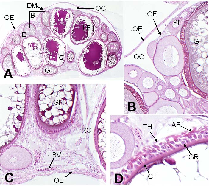
Figure 5-28. (A) Transverse section of adult medaka ovary. (B–D) Magnified images of the ovarian tissues shown in A (positions indicated with boxes). AF, attachment filament; BV, blood vessel; CH, chorion (egg envelope); DM, dorsal mesentery; GE, germinal epithelium; GF, growing follicle; GR, granulosa cell (layer); LF, large follicle; OC, ovarian cavity; OE, ovarian epithelium; PF, primordial follicle; RO, rete ovarii-like structure; TH, theca cells (layer). (Kurokawa et al., 2007. Copyright (2007) National Academy of Sciences, U.S.A.) |