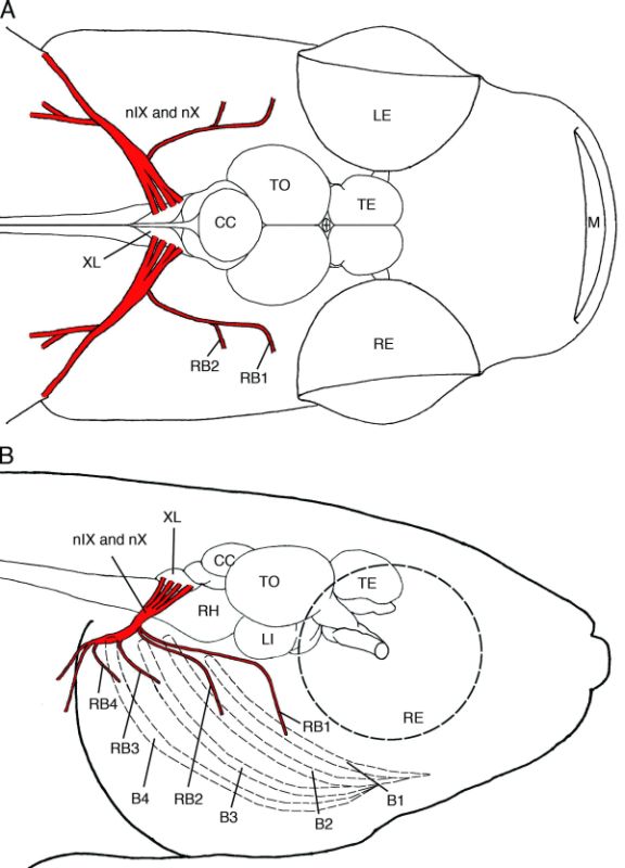
Figure 5-19.
Schematic drawings showing the distribution of the glossopharyngeal and vagus nerves in dorsal view (A) and right lateral view (B). The glossopharyngeal and vagus nerves are shown in red, and the branchial arches are shown as dashed lines. B1, first branchial arch; B2, second branchial arch; B3, third branchial arch; B4, fourth branchial arch; BO, olfactory bulb; CC , corpus cerebelli; LE, left eye; LI, inferior lobe; M, mouth; nⅨ, glossopharyngeal nerve; nⅩ, vagus nerve; RB1, nerve to the first branchial arch; B2, nerve to the second branchial arch; RB3, nerve to the third branchial arch; RB4, nerve to the fourth branchial arch; RE, right eye; RH, rhombencephalon; TE, telencephalon; TO, optic tectum; XL, vagus lobe.
Schematic drawings showing the distribution of the glossopharyngeal and vagus nerves in dorsal view (A) and right lateral view (B). The glossopharyngeal and vagus nerves are shown in red, and the branchial arches are shown as dashed lines. B1, first branchial arch; B2, second branchial arch; B3, third branchial arch; B4, fourth branchial arch; BO, olfactory bulb; CC , corpus cerebelli; LE, left eye; LI, inferior lobe; M, mouth; nⅨ, glossopharyngeal nerve; nⅩ, vagus nerve; RB1, nerve to the first branchial arch; B2, nerve to the second branchial arch; RB3, nerve to the third branchial arch; RB4, nerve to the fourth branchial arch; RE, right eye; RH, rhombencephalon; TE, telencephalon; TO, optic tectum; XL, vagus lobe.