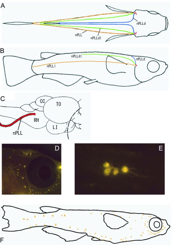
Figure 5-18.
Schematic drawings showing the location of the posterior lateral line nerve in dorsal view (A) and right lateral view (B). The dorsal (blue), dorsolateral (light green), and midbody (orange) branches of the posterior lateral line nerve are depicted. The posterior lateral line nerve (red) exits the brain from the lateral rhombencephalon (C). Neuromasts labeled with fluorescent cytoskeletal markers (DiAsp) in the medaka facial region (see-through strain) (D). Labeled neuromasts at high magnification (E). Schematic drawing showing the distribution of the neuromasts (F).
Schematic drawings showing the location of the posterior lateral line nerve in dorsal view (A) and right lateral view (B). The dorsal (blue), dorsolateral (light green), and midbody (orange) branches of the posterior lateral line nerve are depicted. The posterior lateral line nerve (red) exits the brain from the lateral rhombencephalon (C). Neuromasts labeled with fluorescent cytoskeletal markers (DiAsp) in the medaka facial region (see-through strain) (D). Labeled neuromasts at high magnification (E). Schematic drawing showing the distribution of the neuromasts (F).
CC , corpus cerebelli; LI, inferior lobe; nPLL, posterior lateral line nerve; nPLLd, dorsal ramus of nPLL;
nPLLdl, dorsolateral ramus of nPLL; nPLLl, lateral ramus of nPLL; RH, rhombencephalon; TO, optic tectum.