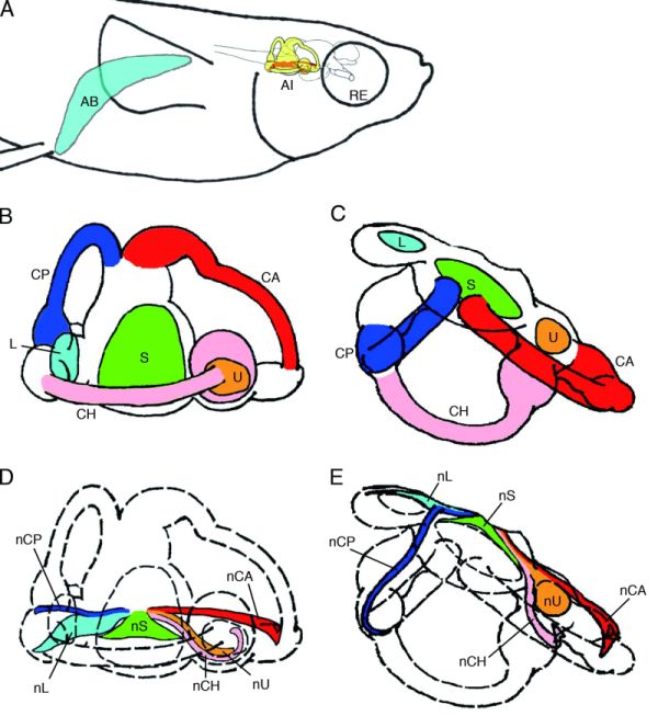
Figure 5-17.
Schematic drawing showing the internal ear (yellow) and air bladder (cyan) in right lateral view (A). Schematic drawings showing the otolith organs and three semicircular canals in right lateral view (B) and dorsal view (C). The saccule (light green), utricle (orange), lagena (cyan), anterior semicircular canal (red), posterior semicircular canal (blue), and horizontal semicircular canal (pink) are depicted. Schematic drawings showing the location of the octaval nerve in right lateral view (D) and dorsal view (E).
Schematic drawing showing the internal ear (yellow) and air bladder (cyan) in right lateral view (A). Schematic drawings showing the otolith organs and three semicircular canals in right lateral view (B) and dorsal view (C). The saccule (light green), utricle (orange), lagena (cyan), anterior semicircular canal (red), posterior semicircular canal (blue), and horizontal semicircular canal (pink) are depicted. Schematic drawings showing the location of the octaval nerve in right lateral view (D) and dorsal view (E).
The branches of the octaval nerve going to the macula of the saccule (light green), the utricle (orange), and the lagena (cyan) are illustrated.
The branches of the octaval nerve going to the ampullary crest of the anterior semicircular canal (red), posterior semicircular canal (blue), and horizontal semicircular canal (pink) are also shown.
AI, internal ear; CA, anterior semicircular canal; CH, horizontal semicircular canal; CP, posterior semicircular canal;
L, otolith of lagena; nCA, nerve to the ampullary crest of CA; nCH, nerve to the ampullary crest of CH;
nCP, nerve to the ampullary crest of CP; nL, nerve to the macula of lagena; nS, nerve to the macula of saccule;
nU, nerve to the macula of utricle; RE, right eye; S, otolith of saccule; U, otolith of utricle.