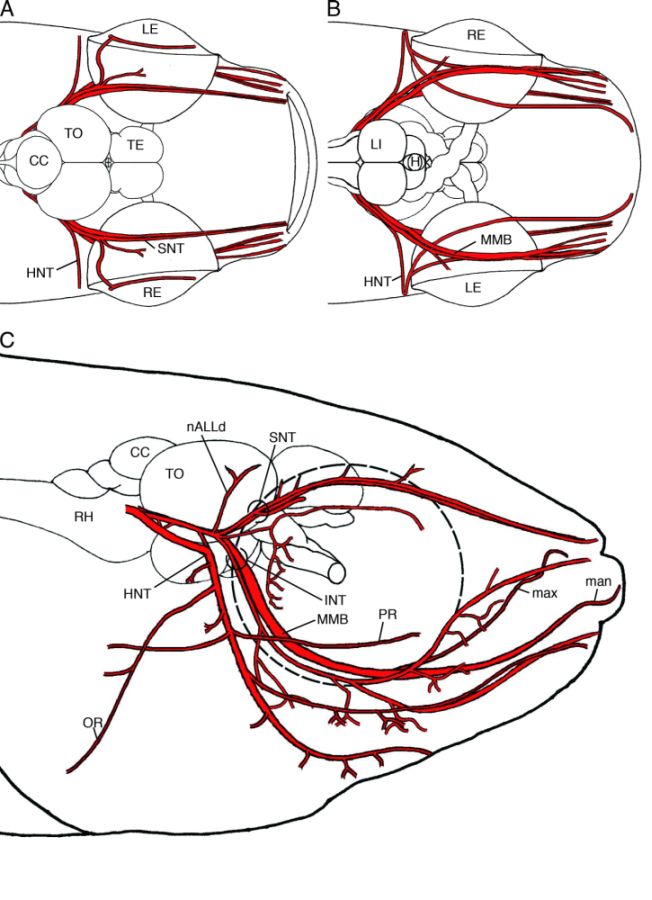
Figure 5-16.
Schematic drawings showing the locations of the trigeminal, facial, and anterior lateral line nerves in dorsal view (A), ventral view (B), and right lateral view (C). All three nerves are shown in red. CC, corpus cerebelli; H, hypophysis ; HNT, hyomandibular nerve trunk; INT, infraorbital nerve trunk; LE, left eye; LI, inferior lobe; man, mandibular ramus; max, maxillary ramus; MMB, maxillomandibular branches; nALLd, dorsal ramus of anterior lateral line nerve; OR, opecular ramus; PR, palatine ramus; RE, right eye; RH, rhombencephalon; SNT, supraorbital nerve trunk; TE, telencephalon; TO, optic tectum.
Schematic drawings showing the locations of the trigeminal, facial, and anterior lateral line nerves in dorsal view (A), ventral view (B), and right lateral view (C). All three nerves are shown in red. CC, corpus cerebelli; H, hypophysis ; HNT, hyomandibular nerve trunk; INT, infraorbital nerve trunk; LE, left eye; LI, inferior lobe; man, mandibular ramus; max, maxillary ramus; MMB, maxillomandibular branches; nALLd, dorsal ramus of anterior lateral line nerve; OR, opecular ramus; PR, palatine ramus; RE, right eye; RH, rhombencephalon; SNT, supraorbital nerve trunk; TE, telencephalon; TO, optic tectum.