|
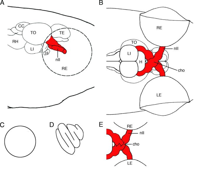
Figure 5-14. Schematic drawings showing the location of the medaka optic nerve in right lateral view (A) and ventral view (B). The optic nerve is shown in red. Schematic drawings of cross sections of the optic nerve of the medaka (C) and the zebrafish (D). Schematic drawing of the optic chiasm of the zebrafish in ventral view (E). CC, corpus cerebelli; cho, optic chiasma; H, hypophysis ; LE, left eye; LI, inferior lobe; nⅡ, optic nerve; RE, right eye; RH, rhombencephalon; TE, telencephalon; TO, optic tectum. |