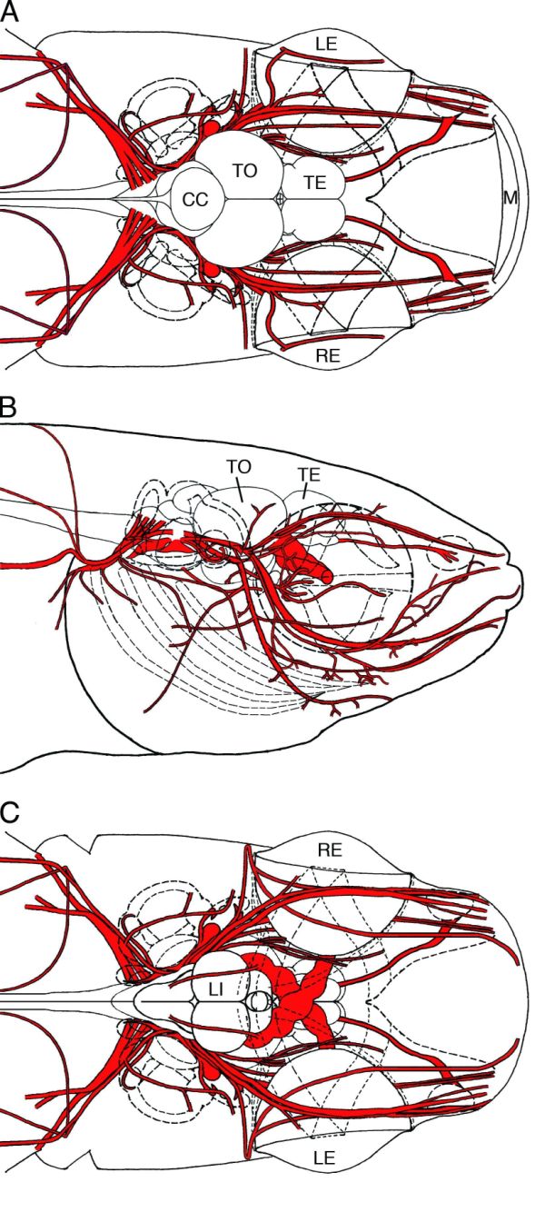
Figure 5-12.
Schematic drawings showing the distribution of the cranial nerves in dorsal view (A), right lateral view (B), and ventral view (C). The cranial nerves are shown in red, and the palatal bone, extraocular muscles, inner ear, and branchial arch are shown in dashed lines. CC, corpus cerebelli; LE, left eye; LI, inferior lobe; M, mouth; RE, right eye; TE, telencephalon; TO, optic tectum.
Schematic drawings showing the distribution of the cranial nerves in dorsal view (A), right lateral view (B), and ventral view (C). The cranial nerves are shown in red, and the palatal bone, extraocular muscles, inner ear, and branchial arch are shown in dashed lines. CC, corpus cerebelli; LE, left eye; LI, inferior lobe; M, mouth; RE, right eye; TE, telencephalon; TO, optic tectum.