|
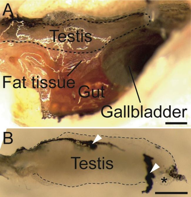
Figure 4-3. Isolation of the testis. (A) Abdominal wall of adult male is cut along the dorsal peritoneum cavity and opened. Testis, fat tissue, gut, and gallbladder are visible in the peritoneum cavity as shown here. (B) Isolated testis. Black peritoneum (arrow head) and fat tissue (asterisk) are attached. Associated tissues of this size have no harmful effects for the rest of the method. The outline of the testes is shown as a dotted line. The lateral views are shown, with the dorsal side toward the top and anterior side toward the right (A, B). Scale bars, 1 mm. |