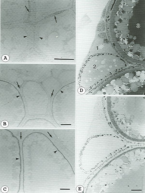
Figure 3-10.
Incorporation of the I125-Choriogenin L into the egg envelope in the ovary. Autograms of unstained sections of the ovary prepared 1 hour (A), 6 hours (B), and 14 hours (C) after the injection. Grains that appeared in the interfollicular space (arrow) 1 hour after the injection (A) disappeared from the section 14 hours after injection (C) (arrow). The inner layer of the oocyte envelope (arrowhead) exhibited a high density of silver grains 6 and 14 hours after the injection. (D, E) Localization of the 125I-Chg. L in the ovary after the injection. Scale bar, 30 μm. (Modified from the Hamazaki et al., 1989.)
Incorporation of the I125-Choriogenin L into the egg envelope in the ovary. Autograms of unstained sections of the ovary prepared 1 hour (A), 6 hours (B), and 14 hours (C) after the injection. Grains that appeared in the interfollicular space (arrow) 1 hour after the injection (A) disappeared from the section 14 hours after injection (C) (arrow). The inner layer of the oocyte envelope (arrowhead) exhibited a high density of silver grains 6 and 14 hours after the injection. (D, E) Localization of the 125I-Chg. L in the ovary after the injection. Scale bar, 30 μm. (Modified from the Hamazaki et al., 1989.)