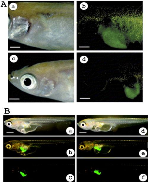
Figure 3-8.
Estrogen-dependent liver-specific expression of GFP in adult male and female of the F1-offspring obtained from one of the chg.L-GFP-emgb/RFP-transgenic medaka strains (A) and in the chg.H-GFP transgenic yolk sac larvae of medaka (B). (A) a–d, the 3-month-old male (a and b) of the F1-offspring derived from one of chg-L1.5 kb/GFP-emgb/RFP-transgenic medaka strains was exposed to 1 ng/ml 17β-estradiol-17 (E2) for 2 days, while the 3-month-old spawning female (c and d) was not exposed to E2. The liver cellular structure was not seen as it was covered by the densely pigmented peritoneum (a and c). However, when observed under the dark field using a GFP filter, the GFP fluorescence was detectable from the outside (b and d). This GFP-positive site corresponded to the liver (b and d). The yellow fluorescent spots found in b and d are pigment cells. The scale bar indicates 1 mm. (B) a–f, fluorescence was observed after exposure of 5.1 nM E2 for 24 hours. The pictures show images of a male (a–c) and a female fish (d–f) with green fluorescence exclusively in the liver. Panels a and d are bright light and panels c and f are fluorescence images. Panels b and e were obtained by the fusing of bright light and fluorescent images. Yellow spots in fluorescence images are caused by auto-fluorescence of pigment cells. Each bar represents 0.2 mm. (Modified from (A) Ueno et al., 2004 and (B) Kurauchi et al., 2005; reprinted with permission from Environ Sci Technol Vol. 39, page 2762–2768. Copyright (2005) American Chemical Society.)
Estrogen-dependent liver-specific expression of GFP in adult male and female of the F1-offspring obtained from one of the chg.L-GFP-emgb/RFP-transgenic medaka strains (A) and in the chg.H-GFP transgenic yolk sac larvae of medaka (B). (A) a–d, the 3-month-old male (a and b) of the F1-offspring derived from one of chg-L1.5 kb/GFP-emgb/RFP-transgenic medaka strains was exposed to 1 ng/ml 17β-estradiol-17 (E2) for 2 days, while the 3-month-old spawning female (c and d) was not exposed to E2. The liver cellular structure was not seen as it was covered by the densely pigmented peritoneum (a and c). However, when observed under the dark field using a GFP filter, the GFP fluorescence was detectable from the outside (b and d). This GFP-positive site corresponded to the liver (b and d). The yellow fluorescent spots found in b and d are pigment cells. The scale bar indicates 1 mm. (B) a–f, fluorescence was observed after exposure of 5.1 nM E2 for 24 hours. The pictures show images of a male (a–c) and a female fish (d–f) with green fluorescence exclusively in the liver. Panels a and d are bright light and panels c and f are fluorescence images. Panels b and e were obtained by the fusing of bright light and fluorescent images. Yellow spots in fluorescence images are caused by auto-fluorescence of pigment cells. Each bar represents 0.2 mm. (Modified from (A) Ueno et al., 2004 and (B) Kurauchi et al., 2005; reprinted with permission from Environ Sci Technol Vol. 39, page 2762–2768. Copyright (2005) American Chemical Society.)