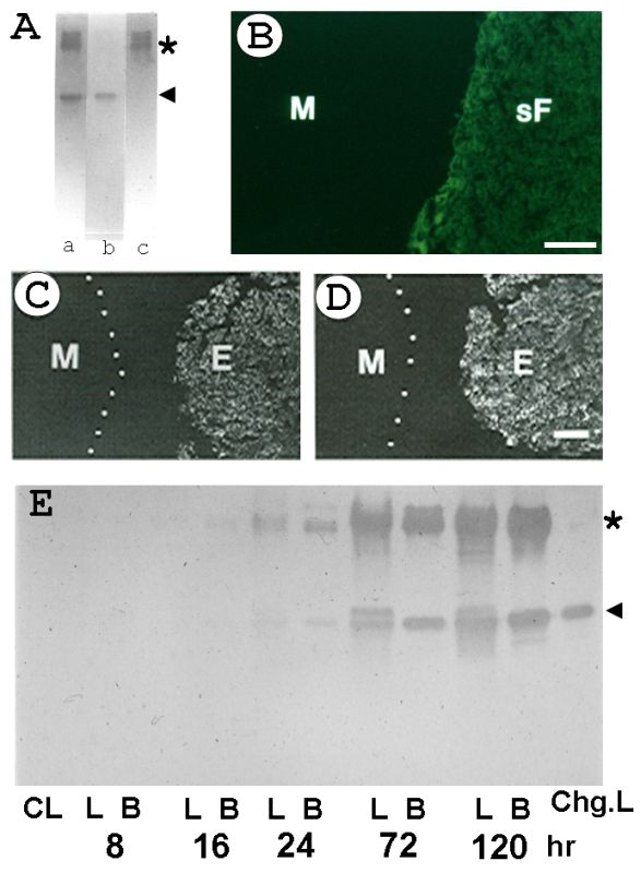
Figure 3-5.
Immunochemical detection of Choriogenins in the liver of spawning medaka female, and estrogen-treated male liver and blood. (A) After the native PAGE, the immunoreactive proteins in the blood of spawning female were detected with anti-medaka egg envelope IgG (a), anti-Choriogenin L IgG (b), and anti-Choriogeini H IgG (c). Asterisk shows the position of Choriogenin H and the arrow indicates the position of Choriogenin L. (B, C, and D) immunohistochemical detection of Choriogenins in the spawning female with anti-medaka egg envelope IgG (B), in the estrogen treated male liver with anti-Choriogenin H IgG (C) and anti-Choriogenin L IgG (D). (E) immunoblotting analysis of the timecourse appearance of Choriogenins in the blood and liver of estrogen-treated male fish with anti-egg envelope IgG. M, normal male liver as the control; sF, spawning female liver; E, estrogen-treated liver; CL, liver from the control (nonestrogenized) male liver; L, B, liver and blood plasma from the estrogenized male fish; chg.L, purified Choriogenin L, the numbers show the time period when the fish was treated in the water containing estrogen (100 ng/ml). (Modified from the Murata et al., 1991, with permission of John Wiley & Sons, Inc. Modified from Murata et al., 1994.)
Immunochemical detection of Choriogenins in the liver of spawning medaka female, and estrogen-treated male liver and blood. (A) After the native PAGE, the immunoreactive proteins in the blood of spawning female were detected with anti-medaka egg envelope IgG (a), anti-Choriogenin L IgG (b), and anti-Choriogeini H IgG (c). Asterisk shows the position of Choriogenin H and the arrow indicates the position of Choriogenin L. (B, C, and D) immunohistochemical detection of Choriogenins in the spawning female with anti-medaka egg envelope IgG (B), in the estrogen treated male liver with anti-Choriogenin H IgG (C) and anti-Choriogenin L IgG (D). (E) immunoblotting analysis of the timecourse appearance of Choriogenins in the blood and liver of estrogen-treated male fish with anti-egg envelope IgG. M, normal male liver as the control; sF, spawning female liver; E, estrogen-treated liver; CL, liver from the control (nonestrogenized) male liver; L, B, liver and blood plasma from the estrogenized male fish; chg.L, purified Choriogenin L, the numbers show the time period when the fish was treated in the water containing estrogen (100 ng/ml). (Modified from the Murata et al., 1991, with permission of John Wiley & Sons, Inc. Modified from Murata et al., 1994.)