|
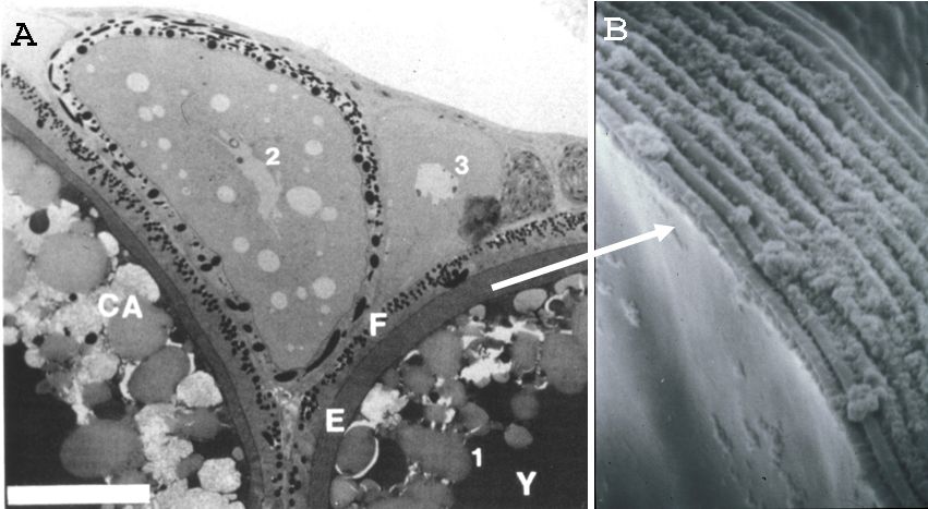
Figure 3-4. Detailed structure of the egg envelope. (A) microscopic observation of the ovarian oocyte (Hamazaki et al., 1989). (B) scanning electron microscopic observation of the egg envelope (Yamagami and Yamamoto, unpublished data). The egg envelope of medaka consists of the outer layer and the inner layer. (Modified from the Hamazaki et al., 1989 (A) and Yamagami and Yamamoto (unpublished data) (B).) |