|
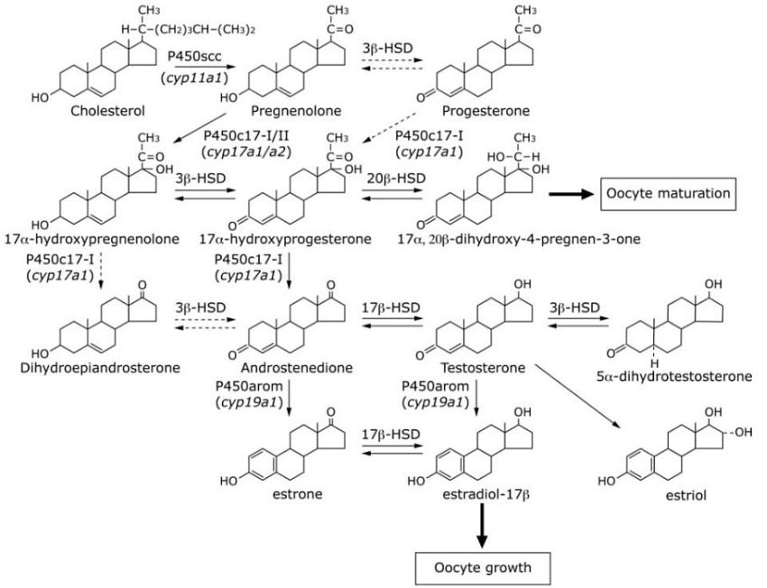
Figure 3-1. Steroidogenic pathways in the medaka ovary. This figure was made on the basis of Kobayashi et al., 1996; Iwamatsu, 1997; Zhou et al., 2007. The solid and dotted lines represent major and minor pathways, respectively. The steroidogenic enzymes (and their genes) that catalyze each pathway are also shown. |